Skin Cancers:
The skin, the largest organ of our body, undertakes many functions. Skin protects the body against possible injuries, prevents fluid loss and helps vitamin D intake. Moreover, skin is protective against both microorganisms and harmful ultraviolet (UV) rays and it helps regulation of the body temperature.
Skin cancer is a malignant growth in skin due to various reasons. Skin cancers may be divided into two main groups, namely melanoma and non-melanoma skin cancers.
Melanoma Skin Cancers
Melanoma is a type of skin cancer that starts in a certain type of cell, called melanocytes that determine color of skin. It is named as the malignant melanoma or cutaneous (originating from skin) melanoma. Melanoma, which is the most malignant type of skin cancers, accounts for approximately 4 percent of all skin cancers. The tumor is generally brown or black in melanoma, as most of the cells continue to produce melanin. However, some melanomas do not produce melanin. In such cases, tumor can appear pink, yellowish brown or even white. Melanoma may develop over congenital or acquired nevi in skin. It may be seen in skin of any body part, including scalp and sole. It is common in neck and back for men and legs, neck and face for women. However, the cancer can also develop in palm, sole or nail bed. In addition, it may occur in eyes, mouth cavity, genitalia or anus in rare cases. While spread of the non-melanoma cancers to other body parts is very rare, melanoma commonly spread to surrounding tissues and other body parts. While melanoma can be treated if detected in early stages, chance of treatment is low in late stages due to its rapid spread.
Other (Non-melanoma) Skin Cancers
Basal cell and squamous cell cancers are the most common tumors of this group. The rarer cases of Merkel cell carcinoma and sebaceous carcinoma are also included in this classification. It is common in parts of the body that are excessively exposed to sunlight such as head, neck and arms. However, this is not a definitive rule.
Basal Cell Carcinoma: It is the most common type of skin cancer. It corresponds to approximately 80 percent of all skin cancers. They appear as small, slowly progressing, bright, pink or red lumps. If left untouched, they tend to cause crust formation, bleeding and ulcerative conversion. Basal cell cancers, which originate from the cells of epidermis - the outermost layer of skin -, grow slowly and spread quite rarely. They are more common in individuals with lighter skin color and colored eyes.
Squamous Cell Carcinoma: It is the second most common type of skin cancers. This cancer originates from the squamous cells of epidermis and it accounts for 16 percent of all skin cancers. It may look like a growing lump or node. It usually has a rough, scaled or crusted surface. In addition, it may show a slowly growing flat formation with red spots.
Actinic keratosis is also called solar keratosis. It is usually assumed a precancerous condition and it occurs due to overexposure to sun. Actinic keratosis generally appears as pink-red or skin colored small, rough or scaled spots. It usually starts on face, ears, dorsum of hands and arms. It may, however, develop in other parts of the body which are exposed to sun. Actinic keratosis is usually characterized with numerous lesions. Some may develop inside the squamous cell carcinoma. Others remain same or progress on their own.
Factors Leading to Skin Cancer
The most critical ones are UVA and UVB rays. All skin cancers, including melanoma, are more common in individuals with long term exposure to ultraviolet rays. Similarly, it has been observed that the risk of melanoma skin cancer is higher in individuals who use solarium frequently for tanning. The lamps that are used in solariums for tanning should bear labels that state “Continuous exposure to UV rays may cause early aging of the skin and skin cancers”. Moreover, it is considered that another label stating “It is necessary to have regular follow-up regarding skin cancer” might be a warning sign for individuals who are continuously exposed to these rays. The nevi in our body are benign tumors; they may also develop during childhood and adolescence periods in addition to the congenital ones (that already exist in birth). Most of the nevi never cause problems. On the other hand, individuals with too many nevi have a greater risk for melanoma. Dysplastic Nevi generally appear similar to ordinary nevi, but they have certain characteristics of melanoma. They are usually larger than other nevi and they have unusual shape and color; most of them never transform into cancer.
It has been identified that the risk of melanoma is 0 to 10 percent for this type of nevi, called Congenital Melanocytic Nevus, depending on size of the nevus. Risk of melanoma is higher for individuals who have a large congenital melanocytic nevus.
Risk of melanoma is 10 times higher for people with white skin and lighter hair color in comparison to those with darker skin tones. Risk of skin cancer increases in people with red and blond hair, white skin, blue or green eye color or the individuals, who have freckles.
It has been shown that the profession of welding and metalwork increases the risk of melanoma in eyes.
UV ray exposure during phototherapy for patients with various skin diseases, such as psoriasis, increases the risk of squamous cell skin cancer.
Individuals with family history of melanoma in one or more first-degree relatives (mother, father, sibling or child) have a higher risk for skin cancer. Approximately 10% of individuals with melanoma have a family member with same disease. The risk may increase due to frequent sun exposure, as members of a family have similar life style, members with light skin color form majority of all family members or both factors may apply. Moreover, gene mutation as a result of any change in genes of the family may be a factor that affects risk of melanoma. A gene mutation is observed in approximately 10 to 40% of the individuals with high rate of melanoma in their family history.
Xeroderma Pigmentosum (XP) is an inherited and rare disease which occurs due to damage to the enzymes that are responsible for repairing DNA in normal circumstances. As the ability of repairing the DNA damaged by sun is lower in individuals with XP, various skin cancers, including melanoma, may develop in body parts that are exposed to sun.
Normal nevi generally appear as brown, skin-colored or black spots on the skin. They may be flat or elevated and erupted, round or ovoid and they measure less than 6 mm in most cases. Nevi may be congenital or develop during childhood and adolescence. Nevi that develop during adulthood should be examined by a physician due to potential risk of a skin cancer. When a nevus develops on the body, its size, shape and color do not change for many years. Some of the nevi may disappear spontaneously over time. Many individuals have nevi and most of these nevi are harmless. However, it is important to recognize any changes in shape, size or color of the nevi due to potential risk of skin cancer.
Symptoms of Skin Cancer
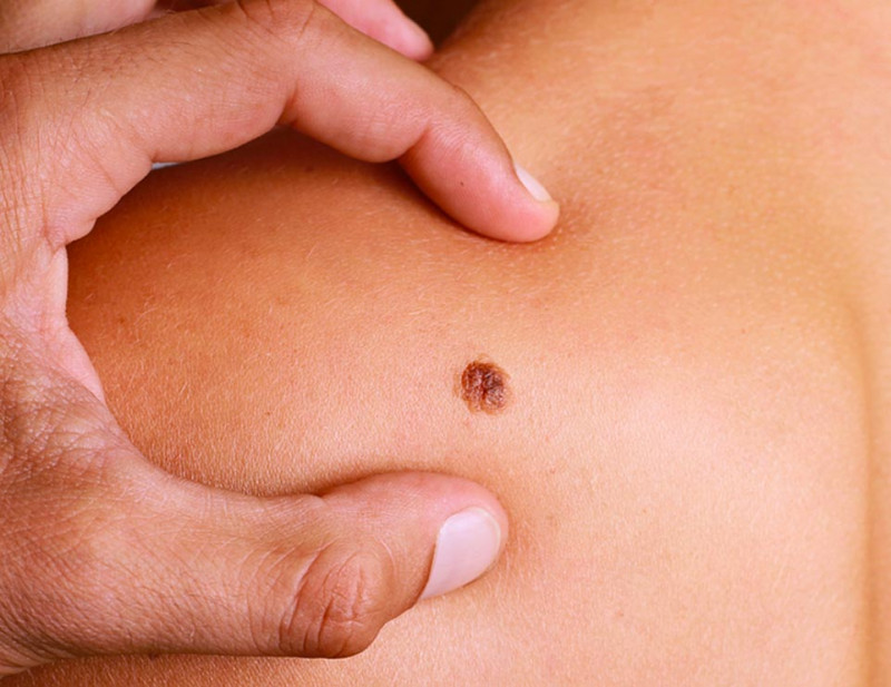
The most critical symptom of skin cancer is a recent-onset spot in skin or any change in size, shape or color in an already existing spot. Other warning signs include persistent wounds, pigmentation beyond the margins of the spot, a recent-onset swelling, itching, tenderness or pain beyond the margins of the spot, any change on surface of the nevi, eruption, bleeding or its appearance in the form of a nodule or lump. It may be challenging to differentiate melanoma from a normal nevus in some cases. When such signs and symptoms are recognized, visiting a physician immediately will ensure that a solution is created quickly for the health problem detected at an early stage.
Self examination of the skin once a month is critical. One should be familiar with his/her body enough to know characteristics of nevi, spots and freckles and to recognize a recent-onset nevus or spot. Self examination of the skin should be performed in a well lightened room in front of a dressing mirror to observe or inspect the entire body. Using a hand mirror may help examining the blind spots of the body, such as back and hip. One out of every 3 melanomas is observed in back of men. Entire body should be checked, including back, palms, soles, scalp, eyes and nails.
Physical examination for skin cancer should be primarily performed by a dermatologist. The spots or nevi on the skin are closely examined with a certain technique, called dermatoscopy. Some skin cancers spread to lymph nodes. In such cases, affected lymph nodes may enlarge and become rougher than they normally are. It is important for individuals with dysplastic nevus syndrome and family history of melanoma to have their skin examined regularly.
Dermatoscopy is the microscopic examination of the skin surface. It is used for diagnosis of nevi and other pigmented lesions. In this method, skin surface is examined with a dermatoscope with a light source that magnifies the images of skin. Nevi were followed up with dermatoscopic images until a few years ago. Next, digital dermatoscope was developed by adding a computer technology to this method. This technique determines punctuate locations by creating a map of the nevi found on skin. Next, dermatoscopic images are obtained and recorded for every single nevus. It is, therefore, possible to compare the images with those that will be obtained in the next examination. Digital dermatoscope also mathematically calculates suspicious changes in nevi and creates an index that shows the risk of malignant melanoma. This index helps diagnosis and treatment planning. While chance of identifying an early-stage melanoma is 60% with naked eye, digital dermatoscope increases this chance to 90 percent.
If skin cancer is suspected for the region of interest, this body part is biopsied and the specimen is examination in laboratory – a procedure called skin biopsy. There are many ways of performing a skin biopsy. Biopsy technique is decided depending on location and size of the suspected area and the type of suspected skin cancer. Depending on condition of the lesion, either incisional biopsy (a part of the lesion is biopsied) or excisional biopsy (the lesion is completely removed) is performed. Additional tests or different treatments may be performed in the light of results. If required, various imaging tests and immunotherapy, targeted therapy and/or radiotherapy can be considered following a surgical procedure that covers the surgical margins sufficiently.
Skin cancers are tumors that are visible to the eye. Similar to other cancers, skin cancer can also be treated before it spreads to other body parts, if relevant changes in skin are identified at early stages. Moreover, side effects of the treatment are minimized, if the skin cancer is treated at an early stage.
Treatment of Skin Cancer
Treatment of skin cancer intends to remove the entire cancerous tissue without leaving any residue. High success rates can be achieved in surgical management. Recurrence of cancer is prevented by removing the cancerous tissue at sufficient dept and width. It is critical to eliminate the cancerous cell and to avoid scar formation due to aesthetic concerns and loss of function in treatment of skin cancer. Patient’s age, overall health, tumor size, skin characteristics and status of spread to lymph glands and internal organs determine the margins and the treatment technique.
Personalized treatment modalities are offered in skin cancer rather than a treatment alone. It is critical that the tumor is removed by a team experienced in treatment of skin cancer.
Radiotherapy may be preferred as the first-line treatment, if it is deemed that the surgical intervention will cause major scarring and the tumor can be managed with radiation with no serious side effect. Non-melanoma skin cancers in cheek, forehead or nose can be successfully treated with radiotherapy, especially if it is a basal cell cancer. Cryosurgery can be used in treatment of small tumors. Local chemotherapy that is applied in the form of a cream or lotion is also considered as an option for superficial skin cancers.
The In Vitro Fertilization implies fertilizing a human oocyte with a human sperm at the laboratory settings and transferring the resultant embryo into the womb.
The robotic rehabilitation is a treatment modality used to restore the walking ability in patients with total or partial loss of the gait function. The robotic rehabilitation is an evidence-based treatment that also involves virtual reality processes.
All pre-and post-operative procedures of the kidney transplant are extremely crucial for the health of both the recipient and the donor.
The check-up examinations allows treatment of the diseases successfully before the condition progresses to the symptomatic stage, as it ensures early diagnosis of many diseases.
An aesthetic look is important for most women. Certain processes that can cause deformities in women's body can make them feel unhappy and desperate.
Breast cancer is the most common type of cancer in women not only in our country but also worldwide.
As coronavirus (COVID-19) pandemic progresses, scientists teach us more about the virus and how it progresses.